
Belajar Bersama Kastrat (BEBEK) 2 raised the theme “Chicken Necropsy,” which was held by PC IMAKAHI UGM under the Department of Strategic Studies on June 1, 2025, at the Macroanatomy Laboratory of the Department of Anatomy, Faculty of Medicine and Health Sciences (FKH) UGM. In its implementation this time, BEBEK 2 collaborated with the Veterinary Integrity and Skill Improvement (VISI) work program of the Department of Human Resource Development, focusing on the theme “The Pulse of Knowledge from the Silent: Provisions for Veterinary Student Career Exploration.” This collaboration aims to integrate theoretical understanding obtained through the VISI seminar with practical experience from the necropsy session organized by BEBEK 2. In addition, necropsy is essential for improving the knowledge of FKH UGM students, enabling them to form qualified prospective veterinarians.
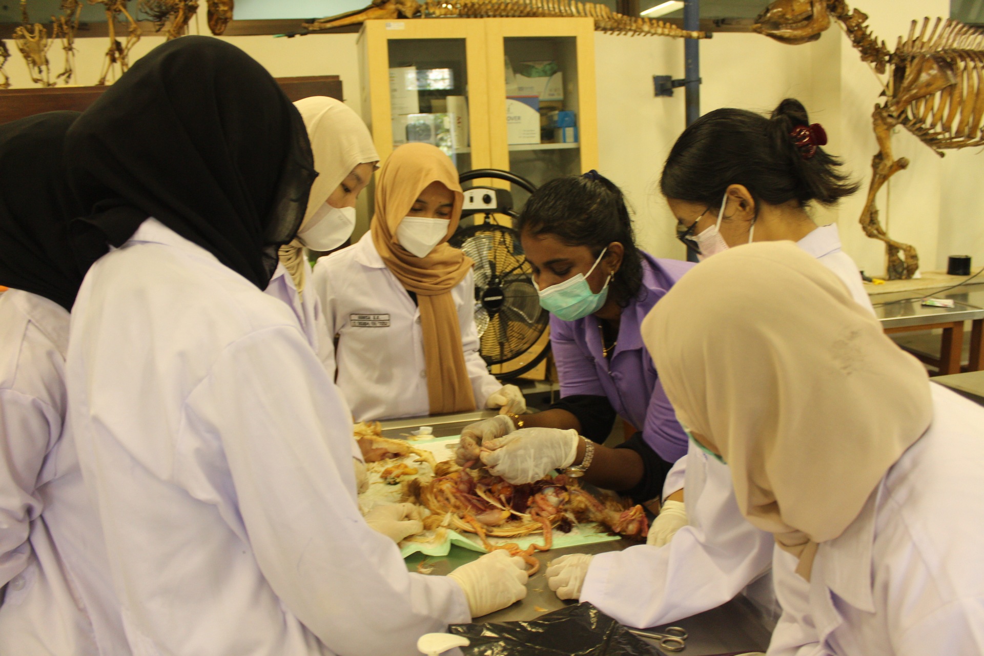
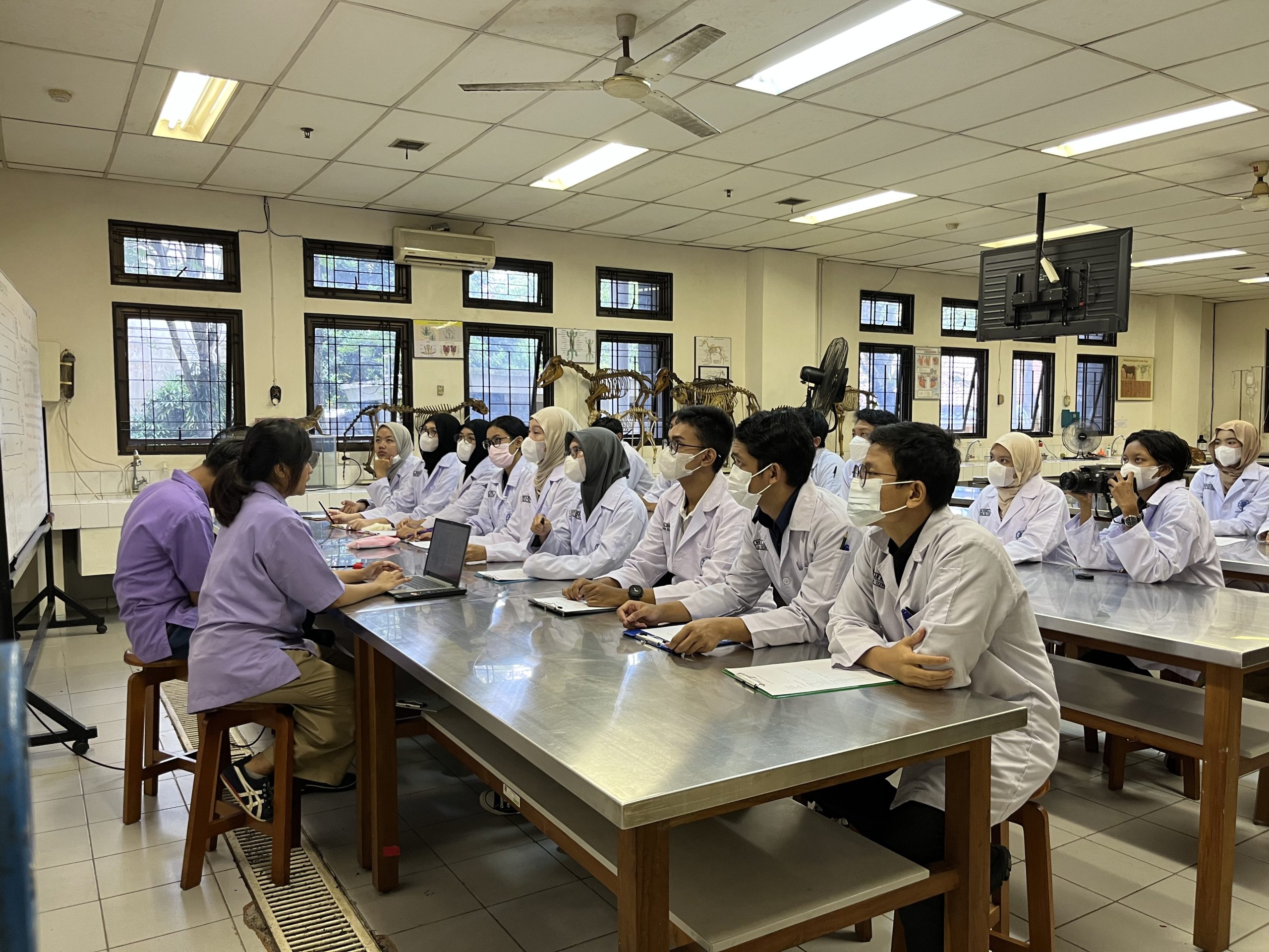
The implementation of BEBEK 2 comprises two main sessions: the material presentation session and the necropsy session. The material presentation session is delivered in the form of a PowerPoint, followed by a necropsy session accompanied by a macroanatomy laboratory assistant. During the necropsy session, participants are divided into four groups, with each group accompanied by a laboratory assistant. Participants participate in interactive necropsy activities, which include external physical examination of the chicken, blood collection, euthanasia using the cardiac embolism technique, and chicken necropsy to identify visceral organs macroanatomically. Chicken blood collection is carried out in the jugular vein, brachial vein, and intracardiac. Meanwhile, euthanasia using the cardiac embolism technique is carried out by inserting air into the chicken’s heart using a syringe while it is still alive. The core of the necropsy session is the necropsy procedure, specifically positioning the chicken in a dorsal position and then making an incision on both the medial thigh and abdomen on both sides of the body. Next, the thigh is pulled to the lateral part, and the incision is continued until the coxofemoral joint. The abdominal skin is cut transversely, then pulled to the anterior part and continued until it reaches the thorax area, extending to the mandible. The abdominal cavity and thorax cavity are opened by making an incision; then the visceral organs are removed for identification. The visceral organs observed in chickens include the digestive system, respiratory system, and reproductive system. Participants identify the visceral organs and record them on the form provided to observe any complications in the chicken’s visceral organs. The BEBEK 2 event was closed with documentation, including all participants, committees, and laboratory assistants.
The BEBEK 2 event has activity objectives that align with several points of the SDGs. First, SDG 3: Good Health and Well-being is related to the detection and control of zoonotic diseases through necropsy practices, which can help create a healthy and prosperous life. Second, SDG 4: Quality Education is highly relevant to this activity because it enhances knowledge and insight, as well as the experience of participants in conducting necropsy practices. This point focuses on improving the quality of education for participants, especially UGM FKH students, to improve the quality of prospective veterinarians. Third, SDG 17: Partnerships for the Goals is realized through collaboration between the Strategic Studies Department and the Human Resources Development Department of PC IMAKAHI UGM.
The necropsy activity of Belajar Bersama Kastrat (BEBEK) 2 is expected to increase knowledge and insight, as well as enhance participant experience through necropsy practices, ultimately improving the quality of qualified prospective veterinarians in the future. Future evaluation of this event is communication that must be improved to avoid miscommunication with several parties.
Cr. Diza Aryani Widhawati
Contact Person Head Organizer: 082243403166 (Diza Aryani Widhawati)
Photo by: Regina Sekar Nathania
